- +1 800 433 4609
- |
- Request Info
Quantitative Sudomotor Axon Reflex Test (QSART)
Introduction
Quantitative Sudomotor Axon Reflex Test (QSART) is a test to evaluate the integrity of the postganglionic sudomotor system along the axon reflex to define the distribution of sweat loss. This is accomplished by the release of bio-impedance electrical stimulation the skin which activates receptors on the eccrine sweat gland. The sweat response is recorded from four sites (forearm and 3 lower extremity sites) and assessed for deficits.
The QSART is a test that measures the autonomic nerves that control sweating. The test is useful in assessing autonomic nervous system disorders, peripheral neuropathies and some types of pain disorders. The test requires a mild electrical stimulation on the skin, which allows to stimulate sweat glands. The QSART measures the volume of sweat produced by this stimulation.
QSART is used to diagnose:
- Painful, small fiber neuropathy when nerve conduction test results are normal
- Disturbances of the autonomic nervous system, which controls the sweat glands, heart, digestive system, other organs, and blood pressure
- Complex pain disorders
- Diabetic neuropathies
- Enzyme disorders
- RSD (Reflex Sympathetic Dystrophy, Complex Regional Pain Syndrome)
- Dysautonomia
- Pharmaceutical agents
- Cosmetics/Consumer goods testing
- Dermatological studies
- Multiple System Atrophy (Shy-Drager syndrome)
Theoretical Background And Methods
The quantitative sudomotor axon reflex test (QSART) has been a routine postganglionic sudomotor function clinical laboratory test since 1983. QSART is a test designed to evaluate the integrity of the postganglionic sympathetic axons. At regional test sites, multicompartmental sweat cells are attached to the limbs. Axon terminals are stimulated by the bio-impedance current, and the sweat response is recorded from a separate compartment. Typically, four regions are studied (one on the forearm and three on the leg).
Functional investigation of thin fibers includes subjective tests, for instance quantitative sensory testing (QST), and objective methods such as measurement of sweat output. Baseline sweat production and responses to electrical stimuli represent functions of postganglionic sympathetic sudomotor neurons. The sweat output can be analyzed after bio-impedance electrical stimulation, recording the induced axon reflex area of sweat production (iodinestarch reaction (Schlereth et al., 2005; Namer et al., 2004; Riedl et al., 1998), or by measuring the total sweat output within this area (quantitative sudomotor axon reflex testing, QSART (Low et al., 1983)). The QSART is widely used to assess impaired sudomotor function clinically in thin fiber neuropathy (Singer et al., 2004). Bio-Impedance stimulation is robust and well-tolerated by the patients. For structural analysis, epidermal nerve fiber and sweat gland density quantified in skin biopsies is used as a diagnostic tool for e.g. diabetic neuropathy (Kennedy et al., 1996; Gibbons et al., 2009). Axonal excitability of sympathetic fibers would be another useful clinical parameter, especially for those diseases in which axonal sodium channels are involved, such as erythromelalgia (Waxman, 2007; Cummins et al., 2004) that is also linked to abnormal sympathetic skin responses (Kazemi et al., 2003).
In order to assess axonal excitability, transcutaneous electrical stimulation was used to induce a combined sudomotor and nociceptor activation. Thereby, both intensity of axon reflex sweating and area of axon reflex erythema can be quantified (Namer et al., 2004). The measurement of axon reflex erythema responses is mainly used under steady state conditions and therefore requires several minutes of electrical stimulation. Thus, systematic analyses of stimulus-response functions for current intensity or frequency would be problematic using this extended protocol. We therefore assessed transient sweat responses and pain to electrical stimulation of 30 s duration. Differential axonal excitability and activation profiles were explored and functionally described for sudomotor neurons and nociceptors by means of current intensity- and frequency-dependent response curves in different body sites.
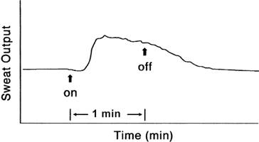
This is a sensitive, reproducible, and quantitative method to test sudomotor function. The basis of the test is that the axon terminal of the sweat gland under the plate electrode is activated by bio-electrical stimulation; the impulse travel centripetally to a branch point and then distally to the axon terminal and a sweating response results. Use of the term "axon reflex" should be discouraged, because only the postganglionic sympathetic sudomotor axon is considered to be involved in this setup. With a latency of 1 to 2 min after the induction of the stimulus, sweat output increases rapidly while stimulation continues; then the stimulator is turned off, and sweat output returns to its prestimulus baseline within 3 min. The area under the curve represents the total amount of sweat output expressed in microliter per square centimeter, and the normal value varies depending on the site of testing, gender, and age of the subject. Distal limbs, male, and younger subjects tend to sweat more. Reduced or absent response indicates postganglionic disorder. Normal response does not rule out preganglionic involvement. Excessive and persistent sweating is also considered abnormal. Comparison is made between the two limbs, and an asymmetry of more than 25% is considered to be abnormal.

