- +1 800 433 4609
- |
- Request Info
Autonomic Nervous System - Introduction
The organs of our body (viscera), such as the heart, intestines and stomach, are regulated by a branch of the nervous system known as the autonomic nervous system. The autonomic nervous system is part of the peripheral nervous system and controls the function of many muscles, glands and organs within the body. We are usually quite unaware of the functioning of our autonomic system because it functions in a reflexive and involuntary manner. For example, we are not aware when our blood vessels change size, and we are (usually) unaware when our hearts speed up or slow down.
What is the Autonomic Nervous System?
The Autonomic Nervous System (ANS) is the involuntary division of the nervous system. It consists of autonomic neurons that conduct impulses from the central nervous system (brain and/or spinal cord) to glands, smooth muscle and cardiac muscle. ANS neurons are responsible for regulating the secretions of certain glands (i.e., salivary glands) and the regulation of heart rate and peristalsis (contraction of smooth muscle in the digestive tract), among other functions
Role of the ANS
The role of the ANS is to constantly fine-tune the functioning of organs and organ systems according to both internal and external stimuli. The ANS helps to maintain homeostasis (internal stability and balance) through the coordination of various activities such as hormone secretion, circulation, respiration, digestion and excretion. The ANS is always "on" and functioning unconsciously, so we are unaware of the important tasks it is performing every waking (and sleeping) minute of every day.
The ANS is divided into two subsystems, the SNS (the sympathetic nervous system) and the PNS (parasympathetic nervous system).
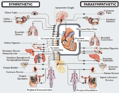
Sympathetic nervous system (SNS) - the SNS triggers what is commonly known as the "fight or flight" response:
- sympathetic neurons are generally considered to belong to the peripheral nervous system, although some of the sympathetic neurons are located in the CNS (central nervous system)
- sympathetic neurons of the CNS (spinal cord) interact with peripheral sympathetic neurons through a series of sympathetic nerve cells bodies known as ganglia
- via chemical synapses within the ganglia, sympathetic neurons join peripheral sympathetic neurons (for this reason, the terms 'presynaptic' and 'postsynaptic' are used to refer to spinal cord sympathetic neurons and peripheral sympathetic neurons, respectively)
- presynaptic sympathetic neurons release acetylcholine at synapses within the sympathetic ganglia. Acetylcholine (Ach) is a chemical messenger that binds nicotinic acetylcholine receptors to the postsynaptic neurons
- postsynaptic neurons release norepinephrine (NE) in response to this stimulus
- prolonged activation of this stimulus response can trigger the release of adrenaline from the adrenal glands (specifically the adrenal medulla)
- once released, NE and adrenaline bind to adrenergic receptors on various tissues, resulting in the characteristic effects of "fight-or-flight"
The following effects are seen as a result of activation of adrenergic receptors:
- increased sweating
- decreased peristalsis
- increased heart rate (increased conduction speed, decreased refractory period)
- pupil dilation
- increased blood pressure (increased contractility, increased ability of the heart to relax and fill)
Parasympathetic nervous system (PNS) - the PNS is sometimes referred to as the "rest and digest" system. In general, the PNS acts in the opposite way to the SNS, reversing the effects of the fight-or-flight response. However, it may be more correct to say that the SNS and the PNS have a complementary relationship, rather than one of opposition.
- the PNS uses Ach as its primary neurotransmitter
- when stimulated, the presynaptic nerve releases acetylcholine (Ach) at the ganglion
- Ach in turn acts on nicotinic receptors of postsynaptic neurons
- postsynaptic nerves then releases acetylcholine to stimulate the muscarinic receptors of the target organ
The following effects are seen as a result of activation of the PNS:
- decreased sweating
- increased peristalsis
- decreased heart rate (decreased conduction speed, increased refractory period)
- pupil constriction
- decreased blood pressure (decreased contractility, decreased ability of the heart to relax and fill)
The Messengers of the SNS and PNS
The autonomic nervous system releases chemical messengers to influence its target organs. The most common are norepinephrine (NE) and acetylcholine (Ach). All presynaptic neurons use Ach as a neurotransmitter. Ach is also released by some sympathetic postsynaptic neurons and all parasympathetic postsynaptic neurons. The SNS uses NE as its principle postsynaptic chemical messenger. NE and Ach are the best-known neurotransmitters of the ANS. In addition to neurotransmitters, certain vasoactive substances are released by postsynaptic automatic neurons, which bind to receptors on target cells and influence the target organ.
How does the SNS mediate its action?
In the sympathetic nervous system, catecholamines (norephinephrine, epinephrine) act on specific receptors located on the cell surface of the target organs. These receptors are called adrenergic receptors.
- Alpha 1 receptors exert their effect on smooth muscle, mainly by constriction. Effects may include constriction of arteries and veins, decreased motility within the GI (gastrointestinal) tract, and constriction of the pupil. Alpa1 receptors are usually located postsynaptically.
- Alpha 2 receptors bind both epinephrine and norepinephrine, thus reducing the effect of alpha 1 receptors to a certain extent. However, alpha 2 receptors have several specific effects of their own, including vasoconstriction. Effects may include coronary artery constriction, constriction of smooth muscle, constriction of veins, decreased intestinal motility and inhibition of insulin release.
- Beta 1 receptors exert their effect mostly on the heart, causing an increase in cardiac output, increased contractility and increased cardiac conduction, leading to an increase in heart rate. There is also stimulation of the salivary glands.
- Beta 2 receptors exert their effect mostly on the skeletal and cardiac muscles. Increased contraction speed and mass of muscles, as well as dilation of blood vessels occurs. Receptors are stimulated by circulating neurotransmitters (catecholamines).
How does the PNS mediate its action?
As mentioned, acetylcholine is the primary neurotransmitter of the PNS. Acetylcholine acts on cholinergic receptors known as muscarinic and nicotinic receptors. Muscarinic receptors exert their effect on the heart. There are two main muscarinic receptors:
M2 receptors- acted on by acetylcholine, M2 receptors are located in the heart; stimulation of these receptors causes the heart to slow (decreased heart rate and contractility and an increase in refractoriness).
M3 receptors- located throughout the body; activation causes increased synthesis of nitric oxide, which results in relaxation of cardiac smooth muscle cells.
How is the autonomic nervous system organized?
As previously discussed, the autonomic nervous system is subdivided into two separate divisions: the sympathetic nervous system and the parasympathetic nervous system. It is important to understand how these two systems function in order to determine how they each affect the body, keeping in mind that both systems work in synergy to maintain homeostasis within the body.
Both the sympathetic and parasympathetic nerves release neurotransmitters, primarily norepinephrine and epinephrine for the sympathetic nervous system, and acetylcholine for the parasympathetic nervous system. These neurotransmitters (also called catecholamines) relay the nerve signals across the gaps (synapses) created when the nerve connects to other nerves, cells or organs. The neurotransmitters then attach to either sympathetic receptor sites or parasympathetic receptor sites on the target organ to exert their effect. This is a simplified version of how the autonomic nervous system functions.
How is the autonomic nervous system controlled?
The ANS is not under conscious control. There are several centers which play a role in control of the ANS:
- Cerebral cortex- the cerebral cortex areas control homeostasis by regulating the SNS, the PNS and the hypothalamus.
- Limbic system- the limbic system is composed of the hypothalamus, the amydala, the hippocampus, and other nearby areas. These structures lie on both sides of the thalamus, just under the cerebrum.
- Hypothalamus- the cells that drive the ANS are located in the lateral medulla. The hypothalamus projects to this area, which includes the parasympathetic vagal nuclei, and also to a group of cells which lead to the sympathetic system in the spinal cord. By interacting with these systems, the hypothalamus controls digestion, heart rate, sweating and other functions.
- Brain stem- the brainstem acts as the link between the spinal cord and the cerebrum. Sensory and motor neurons travel through the brainstem, conveying messages between the brain and spinal cord. The brainstem controls many autonomic functions of the PNS, including respiration, heart rate and blood pressure.
- Spinal cord- two chains of ganglia are located on either side of the spinal cord. The outer chains form the parasympathetic nervous system, while the chains closest to the spinal cord form the sympathetic element.
What are some receptors of the autonomic nervous system?
Sensory neuron dendrites are sensory receptors that are highly specialized, receiving specific types of stimuli. We do not consciously sense impulses from these receptors (except perhaps pain). There are numerous sensory receptors:
- Photoreceptors- respond to light
- Thermoreceptors- respond to alterations in temperature
- Mechanoreceptors- respond to stretch and pressure (blood pressure or touch)
- Chemoreceptors- respond to changes in internal body chemistry (i.e., O2, CO2) and dissolved chemicals during sensations of taste and smell
- Nociceptors- respond to various stimuli associated with damage to tissues (brain interprets the pain)
Autonomic (visceral) motor neurons synapse onto neurons located in the ganglia of the sympathetic and parasympathetic nervous system, which in turn directly innervate muscles and some glands. In this way, visceral motor neurons can be said to indirectly innervate smooth muscles of arteries and cardiac muscle. Autonomic motor neurons work by increasing (in the SNS) or decreasing (in the PNS) activities of their target tissues. In addition, autonomic motor neurons can continue to function even if their nerve supply is damaged, albeit to a lesser extent.
Where are the autonomic nervous system neurons located?
The ANS is essentially comprised of two types of neurons connected in a series. The nucleus of the first neuron is located in the central nervous system. (SNS neurons begin at the thoracic and lumbar areas of the spinal cord, PNS neurons begin at the cranial nerves and sacral spinal cord). The first neuron's axons are located in the autonomic ganglia. In terms of the second neuron, its nucleus is located in the autonomic ganglia, while the axons of the second neuron are located in the target tissue. The two types of giant neurons communicate using acetylcholine. However, the second neuron communicates with target tissue using acetylcholine (PNS) or norepinephrine (SNS). Both the PNS and SNS are connected to the hypothalamus.
| Sympathetic | Parasympathetic | |
| Function | To defend the body against attack | Healing, regeneration and nourishing the body |
| Overall Effect | Catabolic (breaks down the body) | Anabolic (builds up the body) |
| Organs and Glands It Activates | The brain, muscles, the insulin pancreas, and the thyroid and adrenal glands | The liver, kidneys, enzyme pancreas, spleen, stomach, small intestines and colon |
| Hormones and Substances It Increases | Insulin, cortisol and the thyroid hormones | Parathyroid hormone, pancreatic enzymes, bile and other digestive enzymes |
| Body Functions It Activates | Raises blood pressure and blood sugar, and increases heat production | Activates digestion, elimination and the immune system |
| Psychological Qualities | Fear, guilt, sadness, anger, willfulness, and aggressiveness. | Calmness, contentment and relaxation |
| Factors That Activate This System | Stress, fears, anger, worry, excessive thinking and too much exercise | Rest, sleep, meditation, relaxation therapies and feelings of being loved |
Autonomic Nervous System Overview
The autonomic nervous system functions to sustain life by exerting control over the following functions/systems:
- Heart (control of heart rate via contractility, refractory states, cardiac conduction)
- Blood vessels (constriction and dilation of arteries/veins)
- Lungs (relaxation of smooth muscles of the bronchioles)
- Digestive system (gastrointestinal motility, saliva production, sphincter control, insulin production in the pancreas, etcetera)
- Immune system (inhibition of mast cells)
- Fluid balance (constriction of renal artery, rennin secretion)
- Pupil diameter (constriction and dilation of the pupil and ciliary muscle)
- Sweating (stimulates sweat gland secretion)
- Reproductive system (in males, erection and ejaculation; in females, contraction and relaxation of the uterus)
- Urinary system (relaxation and contraction of bladder and detrusor muscles, urethral sphincter)
The ANS, through its two branches (sympathetic and parasympathetic), controls energy expenditure. The sympathetic branch mediates this expenditure while the parasympathetic branch serves a restorative function. In general:
- The sympathetic nervous system causes a speeding up of bodily functions (i.e. heart and respiratory rates) and protect the core by shunting blood from the extremities to the core
- The parasympathetic nervous system causes a slowing of bodily functions (i.e. heart and respiratory rates) and favors healing, rest and restoration, as well as coordinating immune responses
Health can be adversely affected when the effects on one of these systems is unchecked by the other, resulting in a disturbance of homeostasis. The ANS affects changes in the body that are meant to be temporary; in other words, the body should return to its baseline state. It is natural that there should be brief excursions from the homeostatic baseline, but the return to baseline should occur in a timely manner. When one system is persistently activated (increased tone), health may be adversely affected.
The branches of the autonomic system are designed to oppose (and thus balance) each other. For example, as the sympathetic nervous system begins to work, the parasympathetic nervous system goes into action to return the sympathetic nervous system back to its baseline. Therefore, it is not difficult to understand that persistent action by one branch may cause a persistently decreased tone in the other, which can lead to ill health. A balance between the two is both necessary and healthy.
The parasympathetic nervous system has a quicker ability to respond to change than the sympathetic nervous system. Why are we designed this way? Imagine if we weren't: exposure to a stressor causes tachycardia; if the parasympathetic system did not immediately begin to counter the increased heart rate, the heart rate could continue to increase until a dangerous rhythm, such as ventricular fibrillation, developed. Because the parasympathetics are able to respond so quickly, dangerous situations like the one described cannot occur. The parasympathetic nervous system is the first to indicate a change in health condition in the body. The parasympathetics are the main influencing factor on respiratory activity. As for the heart, parasympathetic nerve fibers synapse deep within the heart muscle, while sympathetic nerve fibers synapse on the surface of the heart. Thus, parasympathetics are more sensitive to heart damage.
Transmission of Autonomic Stimuli
Neurons generate and propagate action potentials along their axons. They then transmit signals across a synapse through the release of chemicals called neurotransmitters, which stimulate a reaction in another effector cell or neuron. This process may cause either stimulation or inhibition of the receiving cell, depending which neurotransmitters and receptors are involved.
Propagation- along the axon, axon potential propagation is electrical and occurs through the exchange of N+ and K+ ions across the membrane of the axon. Individual neurons generate the same potential after receiving each stimulus and conduct the axon potential at a fixed rate of velocity along the axon. Velocity is dependent upon the diameter of the axon and how heavily it is myelinated- speed is faster in myelinated fibers because the axon is exposed at regular intervals (nodes of Ranvier). The impulse "jumps" from one node to the next, skipping myelinated sections.
Transmission- transmission is chemical, resulting from the release of specific neurotransmitters from the terminal (nerve ending). These neurotransmitters diffuse across the cleft of the synapse and bind to specific receptors attached to the effector cell or adjoining neuron. Response may be excitatory or inhibitory depending on the receptor. Neurotransmitter-receptor interaction must occur and terminate quickly. This allows for repeated and rapid activation of the receptors. Neurotransmitters can be "reused" in one of three ways:
- Reuptake- neurotransmitters are quickly pumped back into presynaptic nerve terminals
- Destruction- neurotransmitters are destroyed by enzymes located near the receptors
- Diffusion- neurotransmitters may diffuse into the surrounding area and eventually be removed
Receptors- receptors are protein complexes that cover the membrane of the cell. Most interact primarily with postsynaptic receptors; some are located on presynaptic neurons, which allows for finer control of the release of the neurotransmitter. There are two major neurotransmitters in the autonomic nervous system:
- Acetylcholine- the major neurotransmitter of autonomic presynaptic fibers, postsynaptic parasympathetic fibers.
- Norephinephrine- the neurotransmitter of most postsynaptic sympathetic fibers
Functions of the Autonomic Nervous System
The Parasympathetic System
"Rest and digest" response:
- Increase in blood flow to the gastrointestinal tract, which helps to meet the greater metabolic demands placed on the body by the GI tract
- Constriction of the bronchioles when oxygen levels are normalized
- Control of the heart via the Vagus nerve cardiac branches and spinal accessory nerves of the thoracic spinal cord
- Constriction of the pupil allowing for near vision control
- Stimulation of salivary gland production and speeds up peristalsis to aid digestion
- Relaxation/contraction of the uterus and erection/ejaculation in men
In order to understand the functioning of the parasympathetic nervous system, it is helpful to use a real example:
The male sexual response is under direct control of the CNS. Erections are controlled by the parasympathetic system through excitatory pathways. Excitatory signals originate in the brain, through thought, sight or direct stimulation. Regardless of the origin of the excitatory signal, penile nerves respond by releasing acetylcholine and nitric oxide, which in turn signal the smooth muscles of the arteries of the penis to relax and fill with blood. This cascade of events results in erection.
The Sympathetic System
"Fight or Flight" response:
- Stimulation of the sweat glands
- Constriction of peripheral blood vessels to shunt blood to the core, where it is needed
- Increased in supply of blood to skeletal muscles that may be needed for activity
- Dilation of the bronchioles under conditions of low oxygen in the blood
- Reduction in blood flow to the abdomen; decreased peristalsis and digestive activities
- Release of glucose stores from the liver to increase glucose in the bloodstream
As with the parasympathetic system, it is helpful to look at a real example to understand how the sympathetic nervous system functions:
Extreme heat is a stressor that many of us have experienced. When we are exposed to excessive heat, our bodies respond in the following manner: thermal receptors convey stimuli to sympathetic control centers located in the brain. Inhibitory messages are sent along the sympathetic nerves to the blood vessels in the skin, which dilate in response. This dilation of the blood vessels increases the flow of blood to the body's surface so that heat can be lost through radiation from the body surface. In addition to the dilation of blood vessels in the skin, the body also reacts to excessive heat by sweating. This occurs through the rise in body temperature, which is sensed by the hypothalamus, which sends a signal via the sympathetic nerves to the sweat glands, which increase the amount of sweat produced. Heat is lost by evaporation of the sweat produced.
Autonomic Neurons
Neurons that conduct impulses away from the central nervous system are known as efferent (motor) neurons. They differ from somatic motor neurons in that Efferent neurons are not under conscious control. Somatic neurons send axons to skeletal muscle, which is usually under conscious control.
- Visceral efferent neurons- motor neurons whose job it is to conduct impulses to cardiac muscle, smooth muscles and glands. They may originate in the brain or spinal cord (CNS). Two visceral efferent neurons are required to conduct an impulse from the brain or spinal cord to the target tissue.
- Preganglionic (presynaptic) neurons- the cell body of the neuron is located in the grey matter of the spinal cord or brain. It ends in a sympathetic or parasympathetic ganglion.
- Preganglionic autonomic fibers- may begin in the hindbrain, midbrain, upper thoracic spinal cord, or fourth sacral level of the spinal cord. Autonomic ganglia may be found in the head, neck or abdomen. Chains of autonomic ganglia also run parallel to each side of the spinal cord.
- Postganglionic (postsynaptic) neurons- cell body is located in the autonomic ganglion (sympathetic or parasympathetic). The neuron ends in a visceral structure (the target tissue)
Where preganglionic fibers originate and autonomic ganglia are found helps in differentiating between the sympathetic nervous system and the parasympathetic nervous system.
Divisions of the Autonomic Nervous System
A summary of the ANS divisions:
- Consists of visceral (motor) efferent fibers
- Divided into the sympathetic and parasympathetic divisions
- Sympathetic neurons exit the CNS through the spinal nerves located in the lumbar/thoracic regions of the spinal cord
- Parasympathetic neurons exit the CNS via cranial nerves and also spinal nerves located in the sacral spinal cord
- There are always two neurons involved in nerve transmission: presynaptic (preganglionic) and postsynaptic (postganglionic)
- Sympathetic preganglionic neurons are relatively short; postganglionic sympathetic neurons are relatively long
- Parasympathetic preganglionic neurons are relatively long; postganglionic parasympathetic neurons are relatively short
- All neurons of the ANS are either adrenergic or cholinergic
- Cholinergic neurons use acetylcholine (Ach) as their neurotransmitter (including: preganglionic neurons of the SNS and PNS divisions, all postganglionic neurons of the PNS division and postganglionic neurons of the SNS division that act on the sweat glands)
- Adrenergic neurons use norepinephrine (NE) as their neurotransmitter (including all postganglionic SNS neurons except those that act on the sweat glands)
Adrenal Glands
The adrenal glands are located above each kidney (also referred to as the suprarenal glands). They are located at approximately the level of the 12th thoracic vertebrae. The adrenal gland has two parts, an outer cortex and an inner medulla. Both parts produce hormones: the outer cortex produces aldosterone, androgens and cortisol, while the medulla mainly produces epinephrine and norepinephrine. The medulla releases epinephrine and norepinephrine when the body responds to a stressor (i.e., the SNS is activated) directly into the bloodstream.
The cells of the adrenal medulla are derived from the same embryonic tissue as sympathetic postganglionic neurons; therefore the medulla is akin to a modified sympathetic ganglion. The cells of the medulla are innervated by sympathetic preganglionic fibers. In response to neural stimulation, the medulla secretes epinephrine into the bloodstream. Epinephrine effects are similar to norepinephrine.
The hormones produced by the adrenal glands are crucial to normal healthy functioning of the body. Cortisol released as a response to chronic stress (or increased sympathetic tone) can be damaging to the body (i.e., hypertension, altered immune function). If the body is stressed for a prolonged period of time, cortisol levels may be insufficient (adrenal fatigue), causing low blood sugar, excessive tiredness and muscle pain.
Parasympathetic (Craniosacral) Division
The parasympathetic division of the autonomic nervous system is often referred to as the craniosacral division. This is due to the fact that cell bodies of preganglionic neurons are located in the brain stem nuclei, and also in the lateral grey horns of the 2nd through the 4th sacral segments of the spinal cord; hence, the term craniosacral is often used to refer to the parasympathetic division.
Parasympathetic cranial outflow:
- Consists of myelinated preganglionic axons that emerge from the brain stem in cranial nerves (lll, Vll, lX and X)
- Has five components
- Largest is the vagus nerves (X); carry preganglionic fibers comprising nearly 80% of the total outflow
- Axons end in terminal ganglia in the walls of target (effector) organs, where they synapse with ganglionic neurons
Parasympathetic sacral outflow:
- Consists of myelinated preganglionic axons that emerge in the anterior roots of the 2nd through the 4th sacral nerves
- Collectively, they form the pelvic splanchnic nerves, which synapse with ganglionic neurons in the walls of reproductive/elimination organs
Functions of the Autonomic Nervous System
The "3F's" mnemonic (fear, fight, or flight) makes it easy to predict the workings of the sympathetic nervous system. When faced with situations of intense fear, anxiety or stress, the body reacts by speeding up the heart rate, increasing blood flow to vital organs and muscles, slowing digestion, making changes to our vision to allow us to see better and numerous other changes that allow us to react quickly in dangerous or stressful situations. These reactions have allowed us to survive as a species for thousands of years.
As is often the case with the human body, the sympathetic system is perfectly balanced by the parasympathetic division, which returns our system to normal following activation of the sympathetic division. The parasympathetic system not only restores balance, but also performs other important functions in reproduction, rest and sleep, and digestion. Each division uses different neurotransmitters to perform their actions- for the sympathetic nervous system, norepinephrine and epinephrine are the neurotransmitters of choice, while the parasympathetic division uses acetylcholine to perform its duties.
Neurotransmitters of the Autonomic Nervous System
| Neurotransmitters | Sympathetic Nervous System | Parasympathetic Nervous System |
| Acetylcholine | preganglionic fibers | preganglionic fibers; postganglionic fibers at synapses with effector cells (cholinergic) |
| Norepinephrine | postganglionic fibers at synapses with effector cells (adrenergic) |
The above chart describes the major neurotransmitters of the sympathetic and parasympathetic divisions.
There are a few special situations that should be noted:
- Some sympathetic fibers that innervate sweat glands and blood vessels within skeletal muscles release acetylcholine
- Cells of the adrenal medulla are closely related to postganglionic sympathetic neurons; they secrete epinephrine and norepinephrine, similarly to postganglionic sympathetic neurons
Receptors of the ANS
The following chart depicts the receptors of the ANS, including their location:
| Receptors | ANS Division | Location | Adrenergic or Cholinergic |
| Nicotinic receptors | parasympathetic | ANS (both parasympathetic and sympathetic) ganglia; muscle cells | Cholinergic |
| Muscarinic receptors (M2, M3 affect cardiovascular activity) | parasympathetic | M2- located on the heart (acted on by acetylcholine); M3- located on the arterial tree (nitric oxide) | Cholinergic |
| Alpha 1 receptors | sympathetic | mainly located on blood vessels; mainly located postsynaptically | Adrenergic |
| Alpha 2 receptors | sympathetic | located presynaptically on the nerve terminal; also located distal to synaptic cleft | Adrenergic |
| Beta 1 receptors | sympathetic | lipocytes; conduction system of the heart | Adrenergic |
| Beta 2 receptors | sympathetic | mainly located on arteries (coronary and skeletal muscle) | Adrenergic |
Agonist and Antagonist
In order to understand how certain drugs affect the autonomic nervous system, it is necessary to define certain terms:
- Sympathetic agonist (sympathomimetic)- a drug that stimulates the sympathetic nervous system
- Sympathetic antagonist (sympatholytic)- a drug that inhibits the sympathetic nervous system
- Parasympathetic agonist (parasympathomimetic)- a drug that stimulates the parasympathetic nervous system
- Parasympathetic antagonist (parasympatholytic)- a drug that inhibits the parasympathetic nervous system
(One way to keep the terms straight is to think of the suffix -mimetic as meaning "mimic"; in other words, it mimics the action. "Lytic"generally means destruction, so you can think of the suffix -lytic as inhibiting or destroying the action of the system in question).
Responses to Adrenergic Stimulation
Adrenergic responses in the body are stimulated by compounds that are chemically similar to adrenalin. Norepinephrine, which is released from sympathetic nerve endings, and epinephrine (adrenalin) in the bloodstream are the most important adrenergic transmitters. Adrenergic stimulation can have both excitatory and inhibitory effects, depending on the type of receptor on the effector (target) organ:
| Effect on the Target Organ | Stimulatory or Inhibitory Effect |
| Dilation of pupils | stimulatory |
| Decreased secretion of saliva | inhibitory |
| Increase in heart rate | stimulatory |
| Increase in cardiac output | stimulatory |
| Increase in respiratory rate | stimulatory |
| Bronchodilation | inhibitory |
| Increase in blood pressure | stimulatory |
| Decreased motility/secretion of digestive system | inhibitory |
| Contraction of internal rectal sphincter | stimulatory |
| Relaxation of urinary bladder smooth muscles | inhibitory |
| Contraction of internal urethral sphincter | stimulatory |
| Stimulation of lipid breakdown (lipolysis) | stimulatory |
| Stimulation of glycogen breakdown | stimulatory |
Understanding the 3 F's (fear, fight or flight) can help you to imagine the response that can be expected. For example, when faced with a threatening situation, it makes sense that your heart rate and blood pressure will increase, breakdown of glycogen will occur (to provide needed energy) and your rate of respiration will increase. All of these are stimulatory effects. On the other hand, if you are faced with a threatening situation, digestion will not be a priority, thus this function is suppressed (inhibited).
Responses to Cholinergic Stimulation
It is helpful to remember that parasympathetic stimulation is, in general, opposite to the effects of sympathetic stimulation (at least on organs that have dual innervations- there are always exceptions to every rule). An example of an exception is the parasympathetic fibers that innervate the heart- inhibition causes slowing of the heart rate.
| Effect on Target Organ | Stimulatory or Inhibitory Effect |
| Motility/secretion of the digestive system | stimulatory |
| Decrease in heart rate | inhibitory |
| Decreased cardiac output | inhibitory |
| Bronchoconstriction | stimulatory |
| Constriction of pupil | stimulatory |
| Increased salivation | stimulatory |
| Contraction of urinary bladder smooth muscles | stimulatory |
| Relaxation of internal urethral sphincter | stimulatory |
| Contraction of colon muscles (defecation) | stimulatory |
| Relaxation of internal anal sphincter | stimulatory |
Complementary Effects of Both Divisions
The salivary gland is acted upon by both the sympathetic and parasympathetic divisions of the ANS. Sympathetic nerves stimulate constriction of blood vessels throughout the alimentary tract, resulting in decreased blood flow to the salivary glands, which in turn causes thicker saliva. Parasympathetic nerves stimulate the secretion of watery saliva. Thus, the two divisions act differently, but in a complementary fashion.
Cooperative Effects of Both Divisions
Cooperation between the sympathetic and parasympathetic divisions of the ANS can best be seen in the urinary and reproductive systems:
- Reproductive system- sympathetic fibers stimulate ejaculation of semen and reflex peristalsis in females; parasympathetic fibers cause vasodilation, ultimately resulting in erection of the penis in males and the clitoris in females
- Urinary system- sympathetic fibers stimulate the urinary urge reflex by increasing bladder tone; parasympathetic nerves promote contraction of the urinary bladder
Organs Without Dual Innervation
Most organs of the body are innervated by nerve fibers from both the sympathetic and parasympathetic nervous system.
There are a few exceptions:
- adrenal medulla
- sweat glands
- arrector pilli muscles
- most blood vessels
These organs/tissues are only innervated by sympathetic fibers. How does the body regulate their action? The body achieves control through increasing or decreasing of the tone of the sympathetic fibers (firing rate). By controlling the stimulation of sympathetic fibers, the action of these organs can be regulated.
Stress and ANS
When a person is placed in a threatening situation, messages from the sensory nerves are carried to the cerebral cortex and limbic system (the "emotional" brain) and also to the hypothalamus. The anterior portion of the hypothalamus excites the sympathetic nervous system. The medulla oblongata contains centers that control many functions of the digestive, cardiovascular, pulmonary, reproductive and urinary systems. The vagus nerve (which has both sensory and motor fibers) supplies sensory input to these centers through its afferent fibers. The medulla oblongata is itself regulated by the hypothalamus, the cerebral cortex and the limbic system. Thus there are several areas involved in the body's response to stress.
When a person is exposed to extreme stress (picture a terrifying situation that occurs without warning, such as a wild animal poised to attack you), the sympathetic nervous system may become completely paralyzed so that its functions cease completely. The person may be frozen in place, unable to move. They may lose control of their bladder. This is due to an overwhelming number of signals that the brain must "sort" and a corresponding tremendous surge of adrenalin. Thankfully, most of the time we are not exposed to stress of this magnitutude and our autonomic nervous system functions as it should!
Disturbances Clearly Related to Autonomic Involvement
There are numerous diseases/conditions which result from automatic nervous system dysfunction:
- Orthostatic hypotension- symptoms include dizziness/lightheadedness with position change (i.e., going from sitting to standing), fainting, blurred vision, and sometimes nausea. It is sometimes caused by failure of baroreceptors to sense and respond to low blood pressure caused by blood pooling in the legs.
- Horner syndrome- symptoms include decreased sweating, drooping eyelid and pupil constriction affecting one side of the face. It is caused by damage to the sympathetic nerves that supply the eyes and face.
- Hirschsprung's disease- also referred to as congenital megacolon, this disorder features dilation of the colon and severe constipation. It is caused by a lack of parasympathetic ganglia in the wall of the colon.
- Vasovagal syncope- a common cause of fainting, vasovagal syncope occurs when the ANS abnormally responds to a trigger (disturbing sights, straining at stool, standing for prolonged periods) by slowing the heart rate and dilating the blood vessels in the legs, allowing blood to pool in the lower extremities, resulting in a rapid drop in blood pressure.
- Raynaud's phenomenon- this disorder frequently affects young women, causing discoloration of the fingers and toes, and occasionally the ears and other areas of the body. It is caused by extreme vasoconstriction of peripheral blood vessels resulting from hyperactivation of the sympathetic nervous system. It is often precipitated by stress and cold.
- Spinal shock- caused by severe injury or damage to the spinal cord, spinal shock may cause autonomic dysreflexia, characterized by sweating, severe hypertension and loss of bowel or bladder control resulting from sympathetic stimulation below the level of the spinal cord injury that is unchecked by the parasympathetic nervous system.
Autonomic Neuropathy
Autonomic neuropathies are a collection of conditions or diseases that affect sympathetic or parasympathetic neurons (or sometimes both). They may be hereditary (present from birth and passed down from an affected parent) or acquired later in life.
The autonomic nervous system controls many body functions, therefore autonomic neuropathies may cause any number of symptoms and signs that may be elicited through exam or laboratory studies. Sometimes only a single nerve of the ANS is affected; however, physicians must watch for development of symptoms stemming from involvement of other areas of the ANS. Autonomic neuropathies can cause a wide variety of clinical symptoms. These symptoms are dependent upon which nerves of the ANS are affected.
Symptoms may be widely variable and can affect almost all body systems:
- Integumentary system- pale color, lack of ability to sweat affecting one side of the face, itching, hyperalgesia (hypersensitivity of the skin), dry skin, cold feet, brittle nails, worsening of symptoms at night, lack of hair growth on lower legs
- Cardiovascular system- palpitations (racing or skipping beats), tremors, blurring of vision, lightheadedness, presycope, shortness of breath, chest pain, ringing in the ears, lower extremity discomfort, fainting
- Gastrointestinal system- diarrhea or constipation, feeling full after eating very little (early satiety), swallowing difficulties, incontinence, decreased salivation, gastroparesis, fainting during toileting activities, increased gastric motility, vomiting (associated with gastroparesis)
- Genitourinary system- erectile dysfunction, inability to ejaculate, inability to achieve orgasm (in women and men), retrograde ejaculation, urinary urgency and frequency, urinary retention (overflow incontinence), urinary incontinence (stress or urge incontinence), nocturia, enuresis, incomplete emptying of the bladder
- Respiratory system- decreased response to cholinergic stimuli (impaired bronchoconstriction), impaired response to low levels of oxygen in the blood (heart rate and ventilation response)
- Nervous system- burning sensation in the feet, inability to regulate body temperature
- Ocular system- blurring/graying of vision, photophobia, tunnel vision, reduced tearing, difficulty focusing, loss of papillary size over time
Causes of autonomic neuropathy may be related to numerous diseases/conditions, medications used to treat other diseases or procedures (such as surgery):
- Alcoholism- chronic ethanol (alcohol) exposure may lead to impaired axonal transport and damage to cytoskeletal properties. Alcohol has been shown to be toxic to both peripheral and autonomic nerves.
- Amyloidosis- in this condition, insoluble proteins are deposited within various tissues and organs; autonomic dysfunction is common in both primary and hereditary amyloidosis.
- Autoimmune diseases- acute intermittent and variegate porphyria, Holmes-Adie syndrome, Ross syndrome, multiple myeloma and POTS (postural orthostatic tachycardia syndrome) are all examples of diseases that have a known or speculated autoimmune component/cause of the disease. The immune system wrongfully identifies body tissues as foreign and attempts to destroy them, leading to widespread damage to nerves.
- Diabetes-neuropathy occurs commonly in diabetes, affecting both sensory and motor nerves; diabetes is the most common cause of AN.
- Multiple system atrophy-this is a neurological disorder causing degeneration of nerve cells, causing alterations in autonomic functions and problems with movement and balance.
- Nerve damage- nerves may be damaged as a result or trauma or surgery, resulting in autonomic dysfunction.
- Medications-medications used therapeutically to treat other disorders may affect the ANS.
The following are some examples:
- Drugs that increase sympathetic activity (sympathomimetics): amphetamines, monoamine oxidase inhibitors (antidepressants), beta adrenergic stimulants
- Drugs that decrease sympathetic activity (sympatholytics): alpha and beta blockers (i.e. metoprolol), barbiturates, anesthetics
- Drugs that increase parasympathetic activity (parasympathomimetics): anticholinesterases, cholinomimetics, reversible carbamate inhibitors
- Drugs that decrease parasympathetic activity (parasympatholytics): anticholinergics, tranquilizers, antidepressants
Obviously, some individuals cannot control their risk factors for autonomic neuropathy (i.e., hereditary causes of AN). Diabetes is by far the largest contributing factor to AN and puts individuals with the disease at high risk for AN. Diabetics can reduce their risk of AN by controlling their blood sugars carefully to prevent damage to their nerves. Smoking, consuming alcohol regularly, hypertension, hypercholesteremia (high blood cholesterol) and obesity may also increase the risk of developing AN, so these factors should be controlled as much as possible to reduce the risk of developing AN.
Treatment of autonomic dysfunction is largely dependent on the cause of AN. When treatment of the underlying cause is not possible, physicians will attempt various therapies to mitigate symptoms of AN:
- Integumentary system- itching (pruritis) may be treated using medications or may be combated by moisturizing the skin, which may be the primary cause of pruritis; hyperalgesia of the skin may be treated with medications such as gabapentin, a medication used to treat neuropathy and nerve pain.
- Cardiovascular system-symptoms of orthostatic hypotension may be improved by wearing compression stockings, increasing fluid intake, increasing salt in the diet and medications that regulate blood pressure (i.e. fludrocortisones). Tachycardia may be treated with beta blockers. Patients should be counseled to avoid sudden position changes.
- Gastrointestinal system- patients may be counseled to eat small, frequent meals if they have gastroparesis. Medications may sometimes be helpful in increasing motility (i.e., Reglan). Increasing fiber in the diet may help with constipation. Bowel retraining is also sometimes helpful for the treatment of bowel issues. Diarrhea is sometimes helped by antidepressants. Eating a diet that is low in fat and high in fiber may improve digestion and constipation. Diabetic individuals should strive to normalize their blood sugars.
- Genitourinary system- bladder retraining, medications for overactive bladder, intermittent catheterization (used to completely empty the bladder when incomplete bladder emptying is an issue) and medications to treat erectile dysfunction (i.e., Viagra) may be used to treat sexual issues.
- Ocular issues- medications to reduce tearing are sometimes prescribed.
KEY_Web_Disclaimer